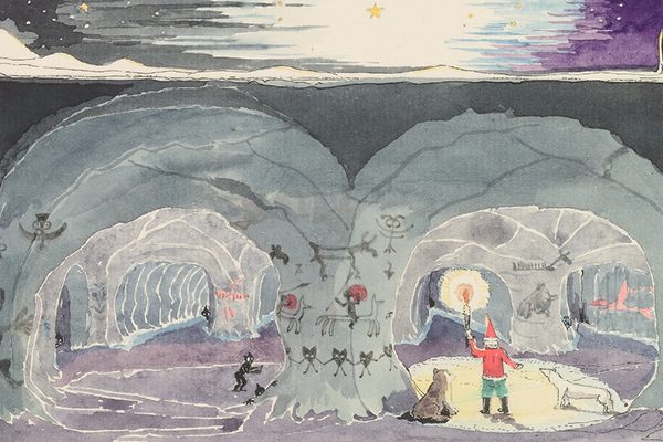
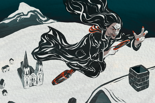
Have a Microscopic Christmas
Thanks to a visualization tool being used to develop stem cell therapies.

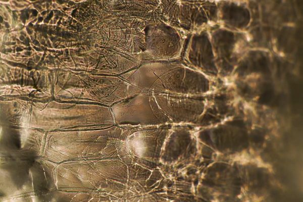
It’s not the world’s smallest Christmas tree, but it’s in the neighborhood. The images above and below are made up of collagen fibers (green) and fat cells (red), distinguished using a laser-based imaging system that might help in the development of stem cell therapies.
The artist of the “cell art” is Catarina Moura, a postdoctoral researcher at the University of Southampton. She works on stem cells—that is, undifferentiated cells that can turn into specialized ones to repair damaged tissue, which has led to their study for a variety of therapies. In Moura’s case, she’s working with a team focused on repairing damaged cartilage. One of the challenges of developing the therapy is monitoring and tracking the cells as they develop into cartilage.

Usually cells are visualized by staining them to glow under fluorescent light. But, as Moura explains, this method interrupts cell development or damages the cells themselves. The system used to make these images, on the other hand, uses ultra-fast lasers to achieve the same effect in a less intrusive way. “Just put the bioengineered cartilage under the microscope and you have the image,” she said in a release. Well, you might also enhance the color to bring out the detail—and add a little holiday spirit.


Follow us on Twitter to get the latest on the world's hidden wonders.
Like us on Facebook to get the latest on the world's hidden wonders.
Follow us on Twitter Like us on Facebook